Remote control of chemical reactions in biological environments could enable a diverse range of medical applications. The ability to release chemotherapy drugs on target in the body, for example, could help bypass the damaging side effects associated with these toxic compounds. With this aim, researchers at California Institute of Technology (Caltech) have created an entirely new drug-delivery system that uses ultrasound to release diagnostic or therapeutic compounds precisely when and where they are needed.
The platform, developed in the labs of Maxwell Robb and Mikhail Shapiro, is based around force-sensitive molecules known as mechanophores that undergo chemical changes when subjected to physical force and release smaller cargo molecules. The mechanical stimulus can be provided via focused ultrasound (FUS), which penetrates deep into biological tissues and can be applied with submillimetre precision. Earlier studies on this method, however, required high acoustic intensities that cause heating and could damage nearby tissue.
To enable the use of lower – and safer – ultrasound intensities, the researchers turned to gas vesicles (GVs), air-filled protein nanostructures that can be used as ultrasound contrast agents. They hypothesized that the GVs could function as acousto-mechanical transducers to focus the ultrasound energy: when exposed to FUS, the GVs undergo cavitation with the resulting energy activating the mechanophore.
“Applying force through ultrasound usually relies on very intense conditions that trigger the implosion of tiny dissolved gas bubbles,” explains co-first author Molly McFadden in a press statement. “Their collapse is the source of mechanical force that activates the mechanophore. The vesicles have heightened sensitivity to ultrasound. Using them, we found the same mechanophore activation can be achieved under much weaker ultrasound.”
Reporting their findings in the Proceedings of the National Academy of Sciences, the researchers demonstrate that this approach can remotely trigger the release of cargo molecules from mechanophore-functionalized polymers using biocompatible FUS.
Drug delivery development
McFadden and colleagues first identified the safe ultrasound parameters for physiological applications. Experiments with 330 kHz FUS revealed a biocompatible upper limit of 1.47 MPa peak negative pressure with a 4.5% duty cycle (3000 cycles per pulse), resulting in an acoustic intensity of 3.6 W/cm2. In a tissue-mimicking gel phantom, these parameters led to a maximum temperature increase of only 3.6 °C.
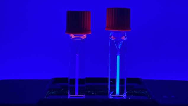
The researchers then investigated whether FUS could activate mechanophore-containing polymers using these biocompatible parameters. They studied the polymer PMSEA containing a chain-centred mechanophore loaded with a fluorogenic small molecule. Exposing a dilute solution of this polymer to biocompatible FUS in the presence of GVs resulted in a strong increase in fluorescence, indicating successful release of the payload – approximately 15% release after 10 min of FUS exposure. Importantly, FUS exposure without the GVs did not trigger a fluorogenic response, confirming that GVs play an essential role as acousto-mechanical transducers.
Next, the researchers examined whether the system was suitable for mechanically triggered drug release. They conjugated the chemotherapy agent camptothecin to the mechanophore followed by polymerization to create PMSEA-CPT, and used FUS to provide controlled release. After 10 min exposure to biocompatible FUS plus GVs, approximately 8% of camptothecin was released. As found for the fluorogenic molecule, no drug release was detected in the absence of GVs.
According to co-first author Yuxing Yao, this is the first time that FUS has been demonstrated to control a specific chemical reaction in a biological setting. “Previously ultrasound has been used to disrupt things or move things,” Yao says. “But now it’s opening this new path for us using mechanochemistry.”
To assess the platform’s future potential for targeted chemotherapy in patients, the researchers investigated its cytotoxicity in vitro on lymphoblast-like Raji cells. Cells incubated for two days with PMSEA-CPT previously exposed to FUS and GVs exhibited a significant decrease in viability. In contrast, no significant cytotoxicity was seen in cells incubated with PMSEA-CPT that had not been exposed to FUS or PMSEA-CPT exposed to FUS but without GVs.

Light-triggered implantable device provides programmable drug delivery
“The mechanically triggered release of molecular payloads from polymers in aqueous media illustrates the power of this approach for non-invasive bioimaging and therapeutic applications of polymer mechanochemistry,” the researchers write. “More broadly, this study demonstrates an approach for achieving remote control of specific chemical reactions under biomedically relevant conditions with the spatiotemporal precision and tissue penetration afforded by FUS.”
Following these initial tests under controlled laboratory conditions, the researchers now plan to test their platform in living organisms. “We are working to translate this fundamental discovery to in vivo applications for drug delivery and other biomedical technologies,” Robb tells Physics World.
- SEO Powered Content & PR Distribution. Get Amplified Today.
- PlatoData.Network Vertical Generative Ai. Empower Yourself. Access Here.
- PlatoAiStream. Web3 Intelligence. Knowledge Amplified. Access Here.
- PlatoESG. Carbon, CleanTech, Energy, Environment, Solar, Waste Management. Access Here.
- PlatoHealth. Biotech and Clinical Trials Intelligence. Access Here.
- Source: https://physicsworld.com/a/biocompatible-focused-ultrasound-delivers-cancer-drugs-on-target/
- :has
- :is
- :not
- :where
- 1
- 10
- 15%
- 3000
- a
- ability
- Academy
- achieved
- achieving
- activating
- Activation
- afforded
- After
- Agent
- agents
- aim
- an
- and
- applications
- applied
- approach
- approximately
- ARE
- around
- AS
- assess
- associated
- At
- author
- based
- BE
- been
- biomedical
- body
- Bound
- broadly
- but
- by
- california
- CAN
- Cancer
- Cause
- Cells
- Changes
- chemical
- Collapse
- colleagues
- conditions
- contains
- contrast
- control
- controlled
- could
- create
- created
- cycle
- cycles
- damaging
- Days
- decrease
- deep
- delivers
- delivery
- demonstrate
- demonstrated
- demonstrates
- detected
- developed
- device
- diagnostic
- DID
- discovery
- Disrupt
- diverse
- drug
- Drugs
- Earlier
- effects
- enable
- energy
- entirely
- environments
- essential
- example
- experiments
- Explains
- exposed
- Exposure
- findings
- First
- first time
- Focus
- focused
- followed
- For
- Force
- found
- from
- function
- fundamental
- future
- GAS
- had
- Have
- heightened
- help
- High
- However
- HTTPS
- identified
- illustrates
- image
- implosion
- importantly
- in
- Increase
- incubated
- information
- initial
- Institute
- into
- issue
- ITS
- jpg
- known
- laboratory
- Labs
- Led
- left
- LIMIT
- living
- lower
- max-width
- maximum
- mechanical
- Media
- medical
- Medical Applications
- method
- min
- molecular
- molecule
- move
- much
- National
- needed
- negative
- New
- no
- now
- of
- on
- only
- opening
- or
- Other
- parameters
- path
- patients
- Peak
- penetration
- per
- phantom
- physical
- Physics
- Physics World
- plan
- platform
- Platforms
- plato
- Plato Data Intelligence
- PlatoData
- Play
- plus
- Polymers
- potential
- power
- precisely
- Precision
- presence
- press
- pressure
- previously
- programmable
- Protein
- provide
- provided
- provides
- pulse
- range
- reaction
- reactions
- release
- released
- relevant
- remote
- required
- researchers
- response
- resulting
- Revealed
- right
- Role
- safe
- safer
- same
- says
- seen
- Sensitivity
- setting
- side
- significant
- small
- smaller
- solution
- Source
- specific
- Statement
- stimulus
- strong
- studied
- studies
- Study
- successful
- suitable
- system
- Target
- targeted
- Technologies
- Technology
- tells
- test
- tests
- that
- The
- The Source
- their
- Them
- then
- These
- they
- things
- this
- Through
- thumbnail
- time
- to
- translate
- trigger
- triggered
- true
- Turned
- two
- under
- undergo
- us
- use
- used
- uses
- using
- usually
- very
- via
- viability
- was
- we
- when
- whether
- which
- with
- within
- without
- working
- world
- write
- zephyrnet


