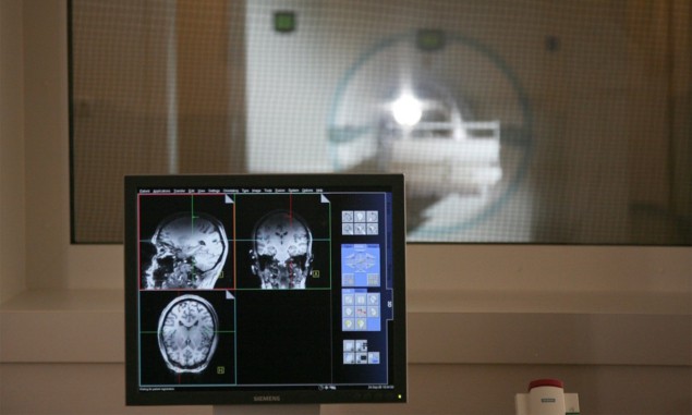
Mapping the uptake of glucose in the brain and body provides clinicians with information about the metabolic dysfunction observed in conditions such as cancer, diabetes and Alzheimer’s disease. This mapping is traditionally performed by administering radioactive substances that act as glucose analogues and can be visualized on medical images.
Scientists know, for example, that tumour cells gobble up glucose more than normal cells. Clinicians exploit this by using 18F-FDG-PET imaging to diagnose and localize tumours and to evaluate treatments. This imaging technique, however, cannot assess downstream metabolites that may be important for diagnosis and treatment evaluation – and it also requires injecting the patient with a radioactive compound.
Another technique, magnetic resonance spectroscopy (MRS) with carbon-13, can quantify downstream metabolites but cannot precisely localize them. Meanwhile, the emerging technique of hyperpolarized 13C-MRS imaging does not provide information about some downstream metabolites, including glutamate and glutamine. Hyperpolarized 13C-MRS imaging also requires injections and uses specialized hardware that may not be available in clinical settings.
Researchers at the Medical University of Vienna have now developed a new approach to mapping glucose metabolism. The technique doesn’t rely on radiation or injections but instead uses clinically-available magnetic resonance imaging (MRI) and oral ingestion of a glucose solution.
2H-MRS
In the researchers’ initial validation study, which appears in Investigative Radiology, participants were imaged with 3 T MRI after fasting overnight and again after ingesting deuterium-tagged glucose solution (deuterium, a stable isotope of hydrogen, is not radioactive). The 2H-MRS scan included a 3D echoless free induction decay sequence, and water suppression was performed using conventional water suppression scheme. After the MRS scan, a 3D T1-weighted magnetization-prepared rapid gradient echo readout scan was performed. An in-house software pipeline was used to process data.
The 2H-MRS imaging approach allowed the researchers to quantify oxidative and anaerobic glucose utilization and assess neurotransmitter synthesis. Yet, they could only measure a limited number of deuterated compounds, and specialized hardware was needed to perform the imaging. So they conducted a follow-up study – now published in Nature Biomedical Engineering – to see whether proton MRS (1H-MRS) at 7 T would provide higher sensitivity, chemical specificity and spatiotemporal resolution than 2H-MRS imaging.
1H-MRS
Studies in animals have shown that deuterium-labelled glucose is readily taken up by brain cells, and deuterons are incorporated into downstream glucose metabolites. Because deuterons substitute protons in the molecule, they do not contribute to the proton spectrum, thus an increase in deuterium-labelled metabolites is reflected by a decrease in metabolite signals in 1H-MRS.
In the 1H-MRS study, five participants (four males and one female) received the deuterium-labelled glucose solution, and their blood glucose levels were measured several times over 90 min. The researchers quantified glutamate, glutamine, γ-aminobutyric acid and glucose deuterated at specific molecular positions. They also mapped deuterated and non-deuterated metabolites. They note that the imaging technique does not require specialized hardware to work with clinically available systems.

Heavy hydrogen tracks glucose metabolism in vivo
Fabian Niess, a research associate involved with the Nature Biomedical Engineering study and lead author of the Investigative Radiology study, explains in a press release that the Investigative Radiology study was “an important step” to demonstrate that the approach worked on lower-field systems “because 3 T MR systems are extremely widespread in clinical applications”.
The researchers conclude that 1H-MRS imaging may facilitate glucose metabolism studies, and they are conducting additional research to verify their approach and preliminary results.
- SEO Powered Content & PR Distribution. Get Amplified Today.
- PlatoData.Network Vertical Generative Ai. Empower Yourself. Access Here.
- PlatoAiStream. Web3 Intelligence. Knowledge Amplified. Access Here.
- PlatoESG. Automotive / EVs, Carbon, CleanTech, Energy, Environment, Solar, Waste Management. Access Here.
- BlockOffsets. Modernizing Environmental Offset Ownership. Access Here.
- Source: https://physicsworld.com/a/mr-spectroscopy-maps-brain-glucose-metabolism-without-requiring-radiation/
- :is
- :not
- $UP
- 3d
- 7
- 8
- a
- About
- AC
- Act
- Additional
- administer
- After
- again
- also
- Alzheimer’s
- an
- and
- animals
- appears
- approach
- ARE
- AS
- assess
- Associate
- At
- author
- available
- BE
- because
- biomedical
- blood
- body
- Brain
- Brain Cells
- but
- by
- CAN
- Cancer
- cannot
- captures
- Cells
- chemical
- Clinical
- clinicians
- Compound
- conclude
- conditions
- conducted
- conducting
- contribute
- conventional
- could
- data
- decrease
- demonstrate
- developed
- Diabetes
- Disease
- do
- does
- Doesn’t
- echo
- emerging
- Engineering
- evaluate
- evaluation
- example
- Explains
- Exploit
- extremely
- facilitate
- female
- For
- four
- Free
- Hardware
- Have
- higher
- However
- HTTPS
- hydrogen
- image
- images
- Imaging
- important
- in
- included
- Including
- Incorporated
- Increase
- information
- initial
- instead
- into
- involved
- issue
- IT
- jpg
- Know
- lead
- levels
- Limited
- mapping
- Maps
- max-width
- May..
- Meanwhile
- measure
- medical
- Metabolism
- method
- min
- molecular
- molecule
- more
- mr
- MRI
- Nature
- Need
- needed
- New
- normal
- now
- number
- of
- on
- ONE
- only
- or
- over
- overnight
- participants
- patient
- Perform
- performed
- Physics
- Physics World
- Pioneering
- pipeline
- plato
- Plato Data Intelligence
- PlatoData
- positions
- precisely
- process
- protons
- provide
- provides
- published
- rapid
- received
- reflected
- rely
- require
- requires
- research
- researchers
- Resolution
- resonance
- Results
- scan
- scheme
- see
- Sensitivity
- Sequence
- settings
- several
- shown
- signals
- So
- Software
- solution
- some
- specialized
- specific
- specificity
- Spectroscopy
- Spectrum
- stable
- studies
- Study
- such
- suppression
- Systems
- taken
- than
- that
- The
- their
- Them
- they
- this
- thumbnail
- times
- to
- traditionally
- treatment
- true
- university
- used
- uses
- using
- validation
- verify
- was
- Water
- were
- whether
- which
- widespread
- with
- without
- Work
- worked
- world
- would
- yet
- zephyrnet


