The resolution of synchrotron X-ray scanning tunnelling microscopy has reached the single-atom limit for the first time, thanks to new work by researchers at Argonne National Laboratory in the US. The advance will have important implications in many areas of science, including medical and environmental research.
“One of the most important applications of X-rays is to characterize materials,” explains study co-leader Saw Wai Hla, Argonne physicist and professor at Ohio University. “Since its discovery 128 years ago by Roentgen, this is the first time that they can be used to characterize samples at the ultimate limit of just one atom.”
Until now, the smallest sample size that could be analysed was an attogram, which is around 10,000 atoms. This is because the X-ray signal produced by a single atom is extremely weak and conventional detectors are not sensitive enough to detect it.
Exciting core-level electrons
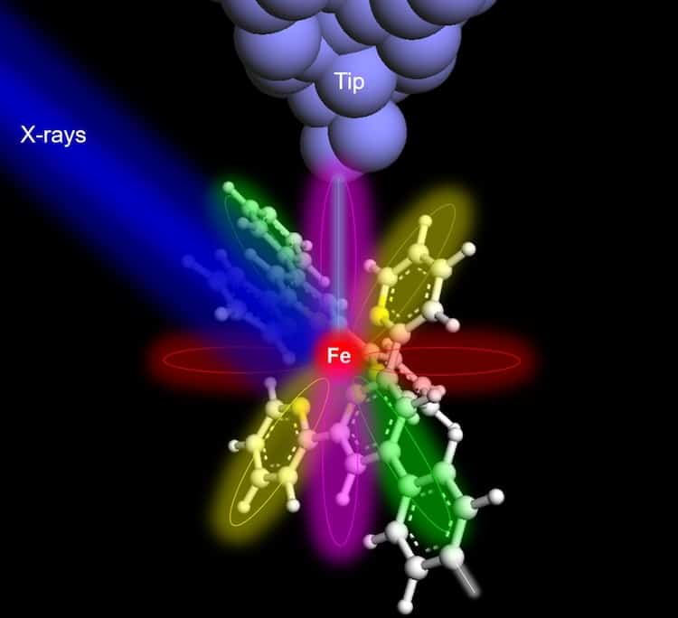
In their work, which the researchers detail in Nature, they added a sharp metallic tip to a conventional X-ray detector to detect X-ray-excited electrons in samples containing iron or terbium atoms. The tip is placed just 1 nm above the sample and the electrons that are excited are core-level electrons – essentially “fingerprints” unique to each element. This technique is known as synchrotron X-ray scanning tunnelling microscopy (SX-STM).

SX-STM combines the ultrahigh-spatial resolution of scanning tunnelling microscopy with the chemical sensitivity provided by X-ray illumination. As the sharp tip is moved across the surface of a sample, electrons tunnel through the space between the tip and the sample, creating a current. The tip detects this current and the microscope transforms it into an image that provides information on the atom under the tip.
“The elemental type, chemical state and even magnetic signatures are encoded in the same signal,” explains Hla, “so if we can record one atom’s X-ray signature, it is possible to extract this information directly.”
Being able to investigate an individual atom and its chemical properties will allow for the design of advanced materials with properties tuned to specific applications, adds study co-leader Volker Rose. “In our work, we looked at molecules containing terbium, which belongs to the family of rare-earth elements, used in applications like electric motors in hybrid and electric vehicles, hard disk drives, high-performance magnets, wind turbine generators, printable electronics and catalysts. The SX-STM technique now provides an avenue to explore these elements without the need to analyse large amounts of material.”

New technique produces colour X-ray images quickly and efficiently
In environmental research, it will now be possible to trace possibly toxic materials down to extremely low levels, adds Hla. “The same is true for medical research where biomolecules responsible for disease could be detected at the atomic limit,” he tells Physics World.
The team says it now wants to explore the magnetic properties of individual atoms for spintronic and quantum applications. “This will impact multiple research fields, from magnetic memory used in data storage devices, quantum sensing and quantum computing to name but a few,” explains Hla.
- SEO Powered Content & PR Distribution. Get Amplified Today.
- PlatoData.Network Vertical Generative Ai. Empower Yourself. Access Here.
- PlatoAiStream. Web3 Intelligence. Knowledge Amplified. Access Here.
- PlatoESG. Automotive / EVs, Carbon, CleanTech, Energy, Environment, Solar, Waste Management. Access Here.
- BlockOffsets. Modernizing Environmental Offset Ownership. Access Here.
- Source: https://physicsworld.com/a/synchrotron-x-rays-image-a-single-atom/
- :has
- :is
- :not
- :where
- 000
- 1
- 10
- 7
- a
- Able
- About
- above
- across
- added
- Adds
- advance
- advanced
- Advanced materials
- ago
- allow
- amounts
- an
- analyse
- and
- applications
- ARE
- areas
- Argonne National Laboratory
- around
- artistic
- AS
- At
- atom
- author
- Avenue
- ball
- BE
- because
- belongs
- between
- but
- by
- CAN
- catalysts
- centre
- characterize
- chemical
- click
- combines
- computing
- conventional
- Core
- could
- created
- Creating
- Current
- data
- data storage
- Design
- detail
- detected
- developed
- Devices
- directly
- discovery
- Disease
- down
- drives
- due
- each
- Electric
- electric vehicles
- Electronics
- electrons
- element
- elements
- enough
- environmental
- essentially
- Even
- excited
- Explains
- explore
- extract
- extremely
- family
- few
- Fields
- First
- first time
- For
- from
- generators
- Green
- Hard
- Have
- he
- high-performance
- How
- HTTPS
- Hybrid
- if
- illuminate
- image
- images
- Impact
- implications
- important
- in
- Including
- individual
- information
- into
- investigate
- issue
- IT
- ITS
- jpg
- just
- just one
- known
- lab
- laboratory
- large
- left
- Level
- levels
- like
- LIMIT
- looked
- Low
- low levels
- Magnets
- many
- material
- materials
- max-width
- medical
- medical research
- Memory
- method
- Microscope
- Microscopy
- molecule
- most
- Motors
- moved
- multiple
- name
- National
- Nature
- Need
- New
- newly
- now
- of
- Ohio
- on
- ONE
- open
- or
- our
- Physics
- Physics World
- plato
- Plato Data Intelligence
- PlatoData
- possible
- possibly
- Produced
- produces
- Professor
- properties
- provide
- provided
- provides
- Quantum
- quantum computing
- quickly
- reached
- record
- Red
- representation
- research
- researchers
- Resolution
- responsible
- right
- same
- saw
- says
- scanning
- Science
- sensitive
- Sensitivity
- sharp
- Signal
- Signatures
- single
- Size
- Space
- specific
- State
- storage
- Study
- Surface
- team
- tells
- Thanks
- that
- The
- their
- then
- These
- they
- this
- Through
- thumbnail
- time
- tip
- to
- Trace
- transforms
- true
- tunnel
- two
- type
- ultimate
- under
- unique
- us
- used
- using
- Vehicles
- via
- wants
- was
- we
- when
- which
- will
- wind
- with
- without
- Work
- world
- x-ray
- years
- zephyrnet


