Magnetic resonance imaging (MRI) is a common medical imaging technique found in hospitals around the world, and something that many of us will experience at some point during our lifetimes. The non-invasive technique identifies diseased tissues by detecting differences in tissue morphology based on the different relaxation times of the tissue after exposure to RF pulses in a magnetic field. Magnetic resonance can also be used as a fundamental measurement mechanism for other types of medical imaging scanners.
There’s an interest in creating portable point-of-care (POC) devices that can image soft tissue just like an MRI scan can. Such systems could rapidly detect aneurysms or fluid pockets, for example, without needing to transport patients to centralized care facilities to perform MRI procedures. The ability to provide this diagnostic information at the bedside with a portable device could improve patient outcomes, reduce the time to treat patients and present lower diagnostic costs for healthcare facilities.
MRI itself is too bulky for bedside imaging, however, and is not suitable for patients who have certain metal implants. Moreover, the power requirements of MRI far outstrip the power capabilities of a portable scanner, as does the weight of the equipment.
These challenges in transferring MRI capabilities to POC devices have led researchers to develop new magnetic resonance-based sensor devices. One such development has come from researchers at Massachusetts Institute of Technology and Harvard University. “Our previous clinical study revealed that skeletal muscle interstitial fluid is an important reservoir for fluid in the body,” lead author Michael Cima tells Physics World. “We needed a magnet design that that could measure that volume at a patient’s bedside.”
POC analysis of muscle tissue
Cima and colleagues chose to create a POC device using a low-field single-sided magnetic resonance (SSMR) sensor to look at skeletal muscle in vivo. Compared with standard MRI equipment, the system is much more portable with a weight of only 11 kg. SSMR sensors use the power of magnetic resonance-based contrast to acquire spectroscopic (non-imaging) data over a limited tissue depth and provide information on the structure of different tissue types – allowing them to be distinguished from one another.
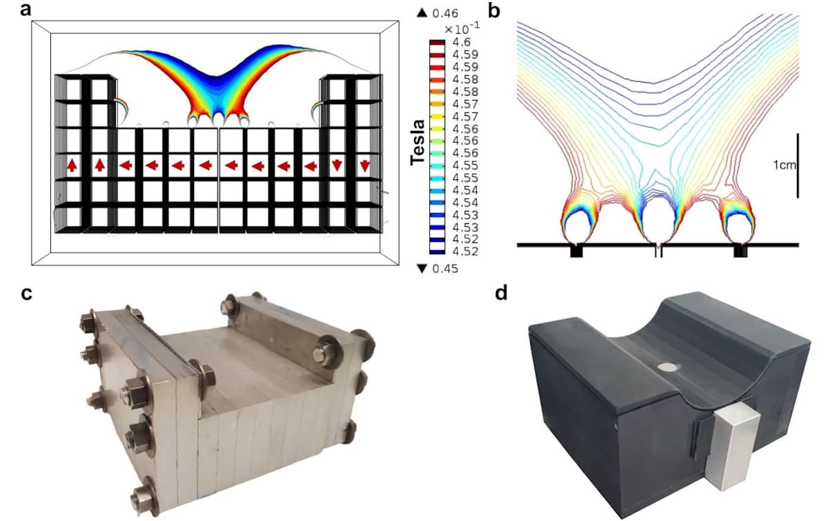
The portable sensor uses a permanent magnet array and surface RF coil to provide low operational power and minimal shielding requirements. The magnet array, constructed from 12.7 mm3 neodymium magnets deployed in aluminium frames, is designed to comfortably seat the calf muscle. The fully assembled sensor with Delrin casing measures 22 × 17.4 x 11 cm.
The sensor can capture low-noise diagnostic measurements within minutes, including T2 relaxation data, which can provide insight into the fluid status, vascular kinetics and oxygenation of skeletal muscle tissue, among other applications. Tissue overheating is avoided by encasing the coil in aluminium nitride, which has a high thermal conductivity that can dissipate generated heat. All these aspects combine to make the SSMR sensor suitable for use as a POC device.
The researchers tested the sensor both in vitro and in vivo, including a clinical study on healthy humans to determine whether the device could successfully detect muscle tissue – which it did. Compared with previous attempts at creating similar SSMR sensors for POC applications, the devices from Cima and his team show better sensitivity and larger penetration depths, and are safer for clinical use.

Portable MR sensor diagnoses dehydration
The new sensor has a penetration depth greater than 8 mm, outperforming other systems described in the literature, which were limited to less than 6 mm depth. Analysis at these levels allowed for an accurate evaluation of the muscle tissue while avoiding signals from other subcutaneous layers, such as the adipose (fat under the skin) tissue that lies closer to the skin’s surface.
The most important outcomes of this study, says Cima, is that “the magnet design met the required performance specifications and is now being used in a 90-patient trial with end-stage renal patients”. When asked about the future potential of these devices, Cima says that “the clinical value of this technology will be demonstrated if we can show that it predicts the ‘dry weight’ [normal weight without excess fluid in the body] of end-stage renal patients. No clinically accepted way to do that currently exists.”
The research is published in Nature Communications.
- SEO Powered Content & PR Distribution. Get Amplified Today.
- PlatoData.Network Vertical Generative Ai. Empower Yourself. Access Here.
- PlatoAiStream. Web3 Intelligence. Knowledge Amplified. Access Here.
- PlatoESG. Carbon, CleanTech, Energy, Environment, Solar, Waste Management. Access Here.
- PlatoHealth. Biotech and Clinical Trials Intelligence. Access Here.
- Source: https://physicsworld.com/a/single-sided-mr-sensor-provides-tissue-analysis-at-the-patient-bedside/
- :has
- :is
- :not
- 10
- 11
- 12
- 17
- 22
- 7
- 8
- a
- ability
- About
- accepted
- accurate
- acquire
- After
- All
- allowed
- Allowing
- also
- among
- an
- analysis
- and
- Another
- applications
- ARE
- around
- Array
- AS
- aspects
- assembled
- At
- Attempts
- author
- avoided
- avoiding
- based
- BE
- being
- Better
- body
- both
- by
- CAN
- capabilities
- capture
- care
- centralized
- certain
- challenges
- chose
- click
- Clinical
- closer
- coil
- colleagues
- combine
- come
- Common
- compared
- constructed
- contrast
- Costs
- could
- create
- Creating
- Currently
- data
- demonstrated
- deployed
- depth
- Depths
- described
- Design
- designed
- detect
- Determine
- develop
- Development
- device
- Devices
- diagnostic
- DID
- differences
- different
- Distinguished
- do
- does
- during
- equipment
- evaluation
- example
- excess
- exists
- experience
- Exposure
- facilities
- far
- Fat
- field
- fluid
- For
- found
- from
- fully
- fundamental
- future
- generated
- greater
- harvard
- harvard university
- Have
- healthcare
- healthy
- High
- his
- hospitals
- However
- HTTPS
- Humans
- identifies
- if
- image
- Imaging
- important
- improve
- in
- Including
- indicate
- information
- insight
- Institute
- interest
- into
- issue
- IT
- itself
- jpg
- just
- larger
- layers
- lead
- Led
- less
- levels
- lies
- like
- Limited
- literature
- Look
- Low
- lower
- magnet array
- Magnetic field
- Magnets
- make
- many
- massachusetts
- Massachusetts Institute of technology
- matching
- max-width
- measure
- measurement
- measurements
- measures
- mechanism
- medical
- met
- metal
- minimal
- Minutes
- MIT
- more
- Moreover
- most
- mr
- MRI
- much
- muscle
- Nature
- needed
- needing
- network
- New
- no
- normal
- now
- of
- on
- ONE
- only
- open
- operational
- or
- Other
- our
- outcomes
- outperforming
- over
- patient
- patients
- penetration
- Perform
- performance
- permanent
- Physics
- Physics World
- plato
- Plato Data Intelligence
- PlatoData
- PoC
- pockets
- Point
- portable
- potential
- power
- Predicts
- present
- previous
- procedures
- Profile
- provide
- provides
- published
- rapidly
- Red
- reduce
- relaxation
- renal
- required
- Requirements
- research
- researchers
- resonance
- Revealed
- safer
- says
- scan
- Sensitivity
- sensor
- sensors
- show
- signals
- similar
- Skin
- Soft
- some
- something
- specifications
- standard
- Status
- structure
- Study
- Successfully
- such
- suitable
- Surface
- system
- Systems
- team
- technique
- Technology
- tells
- tested
- than
- that
- The
- The Future
- the world
- Them
- thermal
- These
- this
- thumbnail
- time
- times
- to
- too
- Transferring
- transport
- treat
- trial
- true
- types
- under
- university
- us
- use
- used
- uses
- using
- value
- volume
- Way..
- we
- weight
- were
- when
- whether
- which
- while
- WHO
- will
- with
- within
- without
- world
- X
- zephyrnet


